GET YOUR QUOTE NOW
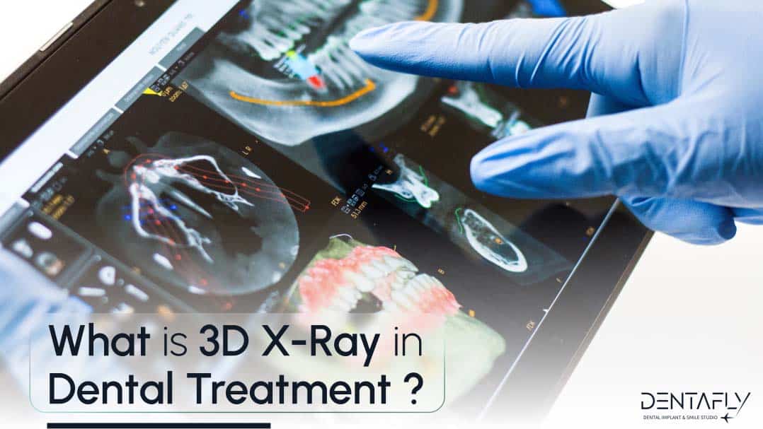
What is 3D X-Ray in dental treatment ?
3D X-ray is the photograph or video of rontgen in 3 dimensional view. 3D X-ray is just like a MRI or a CT Scan that allows dentists and dental implant surgeons to see the inside of your bone to make some calculations, such as:
- Bone level
- Bone density
- Bone length
- Bone width
- Length till the nerve
- Length till the sinus gap
- Whether or not the bone is suitable for immediate loading
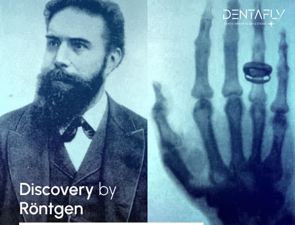
Discovery by Röntgen
On November 8, 1895, German physics professor Wilhelm Röntgen stumbled on X-rays while experimenting with Lenard tubes and Crookes tubes and began studying them. He wrote an initial report “On a new kind of ray: A preliminary communication” and on December 28, 1895, submitted it to Würzburg‘s Physical-Medical Society journal.
This was the first paper written on X-rays. Röntgen referred to the radiation as “X”, to indicate that it was an unknown type of radiation. Some early texts refer to them as Chi-rays having interpreted “X” as the uppercase Greek letter Chi, Χ. The name X-rays stuck, although (over Röntgen’s great objections) many of his colleagues suggested calling them Röntgen rays. They are still referred to as such in many languages, including German, Hungarian, Ukrainian, Danish, Polish, Bulgarian, Swedish, Finnish, Estonian, Slovenian, Turkish, Russian, Latvian, Lithuanian, Japanese, Dutch, Georgian, Hebrew, and Norwegian. Röntgen received the first Nobel Prize in Physics for his discovery.(Wikipedia)
We can say that 3D X-ray is an inseparable part of dentistry, especially for dental implant surgeons. A dentist and a dental implant surgeon can see everything that a naked eye cannot see by looking at the 3D X-ray of a patient. During the planning stage of the patient’s plan, the examination dentist creates the plan for the patient with the dental implant surgeon by checking the 3D X-ray. A certain and healthy plan can be created by calculating the size of the jaw bone. 3D X-ray is very useful for the patient so that they will be comfortable during their treatment.
[ez-toc]
Difference from the 2D Panoramic X-Rays
From a regular 2D Panoramic X-ray, the dentist and the dental implant surgeon can see the teeth, teeth root, whether or not there are any infections, and bone length. The limit of the 2D panoramic X-rays is that the bone length can be deceiving. From a 2D panoramic X-ray, the length can be 9.4 but using the 3D X-ray, we can see the exact place of the nerve canal, and bone width that the dental implant surgeon can actually use to place the implant inside of a strong and reliable jaw bone.
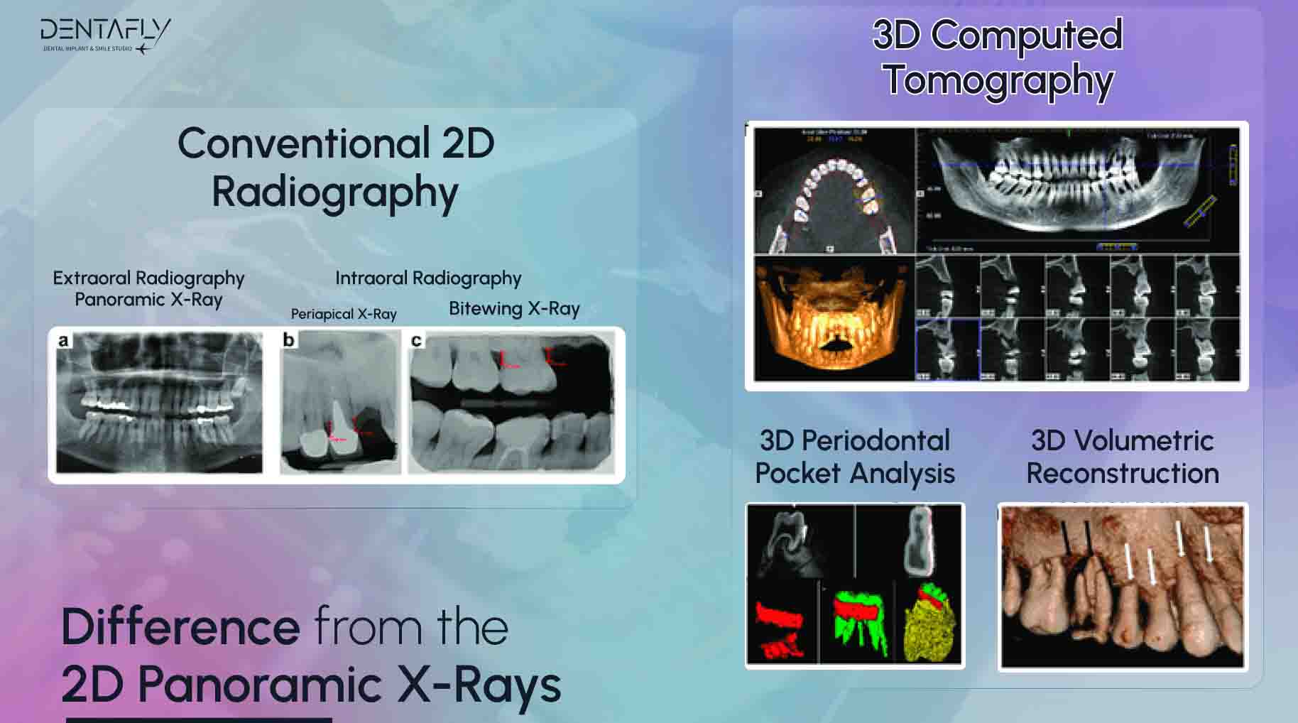
With 2D Panoramic X-rays, we can see whether or not the patient has a cavity, need for root canal treatment, bone loss, hanging sinus gap, and how much the jaw bone is holding the teeth. All of these will help the dentist and the dental implant treatment to create a good and healthy plan that during the procedures the patient will be comfortable and in the future, the treatment will be successful.
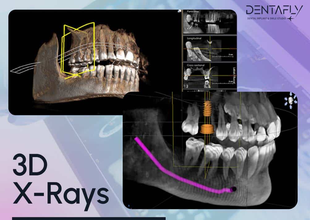
3D X-rays
3D X-ray, unlike 2D X-rays, is the three-dimensional x-ray imaging. Which basically creates the entire oral cavity by designing a 3D image. The 3D image can be moved to all directions so that the dentist and the implant surgeon can move the oral cavity 3D image to see the bone width, bone length, bone shape, bone density, and the nerve canal. Using the 3D X-ray method is the best way to create the best treatment plan.
3D X-rays can be used to create many different treatment plans and types for each and every one of the patients individually. Different treatment plans and types that can be created by 3D x-Ray are:
- Orthodontic procedures
- Planning dental implant placement for the damaged teeth
- Diagnosis of the temporomandibular joint dysfunction
- Finding the strong slice of the bone for placement of the implant(s)
- Locating the origin of pain or a disease
- Finding, measuring, and treating jaw bone tumours
- Collecting data for the bone structure and health
- Reconstructive surgery for the jaw and dental implants
- Planning for root canal treatment, sinus lift, and bone graft
Is 2D panoramic X-ray or 3D X-ray better for planning?
Mostly dental clinics use 2D X-Ray machines but 3D X-Ray is an union of 2D and plus. 3D X-Ray gives better definitions nowadays but It was blurred in the first days of 3D technologies.
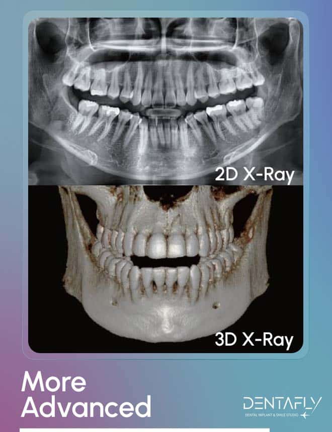
More advanced
3D X-ray is far better and advanced than 2D panoramic X-ray. 3D X-ray can see the infection, bone length, bone density, bone width, and bone quality much better than a 2D panoramic X-ray. 3D X-ray can show slices from the jaw bone that will make the panning process much more certain and the results of the dental implant treatment will be much more predictable.
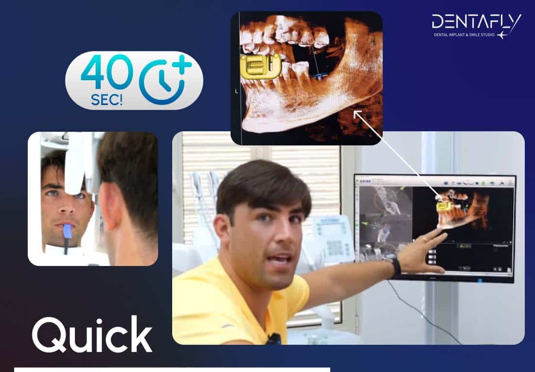
Quick
Even though the 3D X-ray is far more capable than the 2D panoramic X-ray, it takes about 40 seconds to complete from start to the end. The radiology specialist will accompany you and prepare you for the 3D X-ray and 2D panoramic X-ray. The radiology specialist will explain the process to you and within approximately 40 seconds, the 3D X-ray is taken.
Better diagnosis and planning
The dentist and the dental implant surgeon will be able to see each slice of your upper and lower jaw bone to find the good quality bone to place the dental implant or dental implants and the patients will be more relaxed to know that they are in good hands.
Reduced risk of diagnosing and planning
The dentist and the dental implant surgeon will be able to see the front, back, sides, and the insides of the jaw bone of the patient reduces the risk of the dental implant treatment from the beginning. The dental implant surgeon will be able to see the bone quality beforehand thus, the dental implant surgeon will make the correct choice of the placement of the dental implant or dental implants.
Better image
3D X-ray can show the inside of the oral cavity, inside of the jaw bone, and by using the computer programmes, the dentist and the dental implant surgeon can use different filters to make calculations. With 2D panoramic X-ray, the dentist and the dental implant surgeon can play with the contrast and the colour but the technology of 2D panoramic X-ray is far behind a 3D X-ray.
Better explanation
With 3D X-ray, it is much easier to explain to the patient about their treatment plan. The dentist and the dental implant surgeon will walk the patients through their plan so that the patient does not have any question in their minds. If the patients have a question they would like to ask about their treatments, the dentist and the dental implant surgeon will explain everything using the 3D X-ray image.
2D X-Rays Versus 3D X-Rays
| 2D | VERSUS | 3D | |||
| Versatile imaging system | ✔ | ||||
| Bone length, width, quality, density calculation | ✔ | ||||
| 3 Dimensional view | ✔ | ||||
| ✔ | Infection, cyst, etc imaging | ✔ | |||
| ✔ | Calculating sinus gap | ✔ | |||
| ✔ | Location of the nerves | ✔ | |||
| ✔ | Imaging teeth roots inside the bone | ✔ | |||
As it is seen, 2D X-ray can show infection, sinus, nerves, and the jaw bone that hold the roots of the teeth. 2D X-ray shows these images like a photograph, on the other hand, with 3D X-ray, you can move the jaw of the patient on the computer and check the insides outsides of the bone so that there is no surprise during the operation. From 2D, you cannot check the width of the bone and sometimes it can be misleading trusting 2D X-ray. Calculations made of sinus and nerves are not always correct on 2D X-ray. The calculations may show enough place for a dental implant but on 3D X-ray, it is seen that the top part of the bone is actually empty on the inside.
In conclusion
3D X-ray gives far better and more accurate images of the patients’ oral cavity and their bone quality that a 2D panoramic X-ray cannot. Not every clinic has a 3D X-ray machine within their facility. Here, at Dentafly, we are using the best 3D X-ray machine in the world. We are caring about our patients and we have been using the highest quality and lowers radiation emitting 3D X-ray and 2D panoramic X-ray machines.
Reference(s)


Leave a Reply